

-
Rapid developments in live-cell three-dimensional (3D) microscopy enable imaging of cell morphology and signaling with unprecedented detail. However, tools to systematically measure and visualize the intricate relationships between intracellular signaling, cytoskeletal organization and downstream cell morphological outputs do not exist. Here, we introduce u-shape3D, a computer graphics and machine-learning pipeline to probe molecular mechanisms underlying 3D cell morphogenesis and to test the intriguing possibility that morphogenesis itself affects intracellular signaling. We demonstrate a generic morphological motif detector that automatically finds lamellipodia, filopodia, blebs and other motifs. Combining motif detection with molecular localization, we measure the differential association of PIP2 and KrasV12 with blebs. Both signals associate with bleb edges, as expected for membrane-localized proteins, but only PIP2 is enhanced on blebs. This indicates that subcellular signaling processes are differentially modulated by local morphological motifs. Overall, our computational workflow enables the objective, 3D analysis of the coupling of cell shape and signaling.

First images of antibody attacking neuron receptor are published
-
Autoimmune encephalitis occurs when antibodies or T-cells go rogue and attack the brain. In this study, UTSW researchers and colleagues from Berlin used cryo-electron microscopy to determine the atomic structures of autoantibodies bound to the GABA-A receptor. That receptor is an important protein in the brain and a target in autoimmune encephalitis.
-
Autoantibodies targeting neuronal membrane proteins can cause encephalitis, seizures, and severe behavioral abnormalities. While antibodies for several neuronal targets have been identified, structural details on how they regulate function are unknown. Here, we determined cryo-electron microscopy structures of antibodies derived from an encephalitis patient bound to the γ-aminobutyric acid type A (GABA-A) receptor. These antibodies induced severe encephalitis by directly inhibiting GABA-A function, resulting in nervous system hyperexcitability. The structures reveal mechanisms of GABA-A inhibition and pathology. One antibody directly competes with a neurotransmitter and locks the receptor in a resting-like state. The second antibody targets the subunit interface involved in binding benzodiazepines and antagonizes diazepam potentiation. We identify key residues in these antibodies involved in specificity and affinity and confirm structure-based hypotheses for functional effects using electrophysiology. Together these studies define mechanisms of direct functional antagonism of neurotransmission underlying autoimmune encephalitis in a human patient.
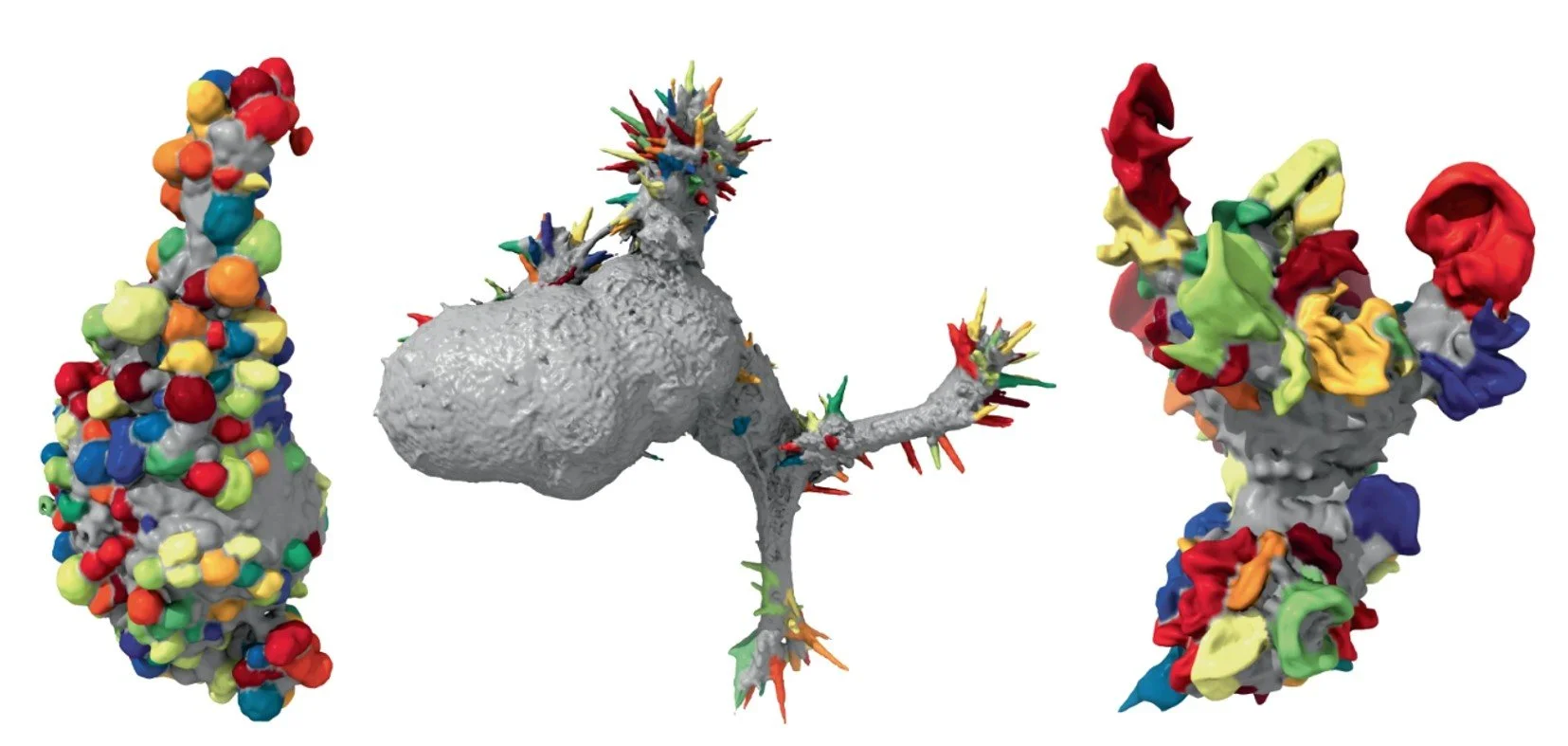
Live cell 3D microscopy reveals links between cell shape and signaling
-
Rapid developments in live-cell three-dimensional (3D) microscopy enable imaging of cell morphology and signaling with unprecedented detail. However, tools to systematically measure and visualize the intricate relationships between intracellular signaling, cytoskeletal organization and downstream cell morphological outputs do not exist. Here, we introduce u-shape3D, a computer graphics and machine-learning pipeline to probe molecular mechanisms underlying 3D cell morphogenesis and to test the intriguing possibility that morphogenesis itself affects intracellular signaling. We demonstrate a generic morphological motif detector that automatically finds lamellipodia, filopodia, blebs and other motifs. Combining motif detection with molecular localization, we measure the differential association of PIP2 and KrasV12 with blebs. Both signals associate with bleb edges, as expected for membrane-localized proteins, but only PIP2 is enhanced on blebs. This indicates that subcellular signaling processes are differentially modulated by local morphological motifs. Overall, our computational workflow enables the objective, 3D analysis of the coupling of cell shape and signaling.

Artificial intelligence tools like sequence modeling algorithms enable proactive solutions to diseases caused by genetic mutations
-
Newly developed artificial intelligence (AI) programs accurately predicted the role of DNA’s regulatory elements and three-dimensional (3D) structure based solely on its raw sequence, according to two recent studies in Nature Genetics. These tools could eventually shed new light on how genetic mutations lead to disease and could lead to new understanding of how genetic sequence influences the spatial organization and function of chromosomal DNA in the nucleus.
-
Epigenomic profiling has enabled large-scale identification of regulatory elements, yet we still lack a systematic mapping from any sequence or variant to regulatory activities. We address this challenge with Sei, a framework for integrating human genetics data with sequence information to discover the regulatory basis of traits and diseases. Sei learns a vocabulary of regulatory activities, called sequence classes, using a deep learning model that predicts 21,907 chromatin profiles across >1,300 cell lines and tissues. Sequence classes provide a global classification and quantification of sequence and variant effects based on diverse regulatory activities, such as cell type-specific enhancer functions. These predictions are supported by tissue-specific expression, expression quantitative trait loci and evolutionary constraint data. Furthermore, sequence classes enable characterization of the tissue-specific, regulatory architecture of complex traits and generate mechanistic hypotheses for individual regulatory pathogenic mutations. We provide Sei as a resource to elucidate the regulatory basis of human health and disease.
-
To learn how genomic sequence influences multiscale three-dimensional (3D) genome architecture, this manuscript presents a sequence-based deep-learning approach, Orca, that predicts directly from sequence the 3D genome architecture from kilobase to whole-chromosome scale. Orca captures the sequence dependencies of structures including chromatin compartments and topologically associating domains, as well as diverse types of interactions from CTCF-mediated to enhancer–promoter interactions and Polycomb-mediated interactions with cell-type specificity. Orca enables various applications including predicting structural variant effects on multiscale genome organization and it recapitulated effects of experimentally studied variants at varying sizes (300 bp to 90 Mb). Moreover, Orca enables in silico virtual screens to probe the sequence basis of 3D genome organization at different scales. At the submegabase scale, it predicted specific transcription factor motifs underlying cell-type-specific genome interactions. At the compartment scale, virtual screens of sequence activities suggest a model for the sequence basis of chromatin compartments with a prominent role of transcription start sites.
Lead Researchers: Colleen Noviello, Ph.D. & Ryan Hibbs, Ph.D.
Lead Researchers: Meghan Driscoll, Ph.D., Gaudenz Danuser, Ph.D., Kevin Dean, Ph.D., Reto Fiolka, Ph.D.
Lead Researcher: Jian Zhou, Ph.D.
Research enabled by BioHPC
“For all our years in Dallas, BioHPC has been the pacemaker for my lab’s discoveries of how metastatic cells survive and proliferate”
“BioHPC has addressed all our computing needs since the start of my lab, and really enabled us to focus on the science of genome regulation using state-of-the-art computing.”